BHU inaugurates fetal echocardiography machine and advanced cardiac imaging lab
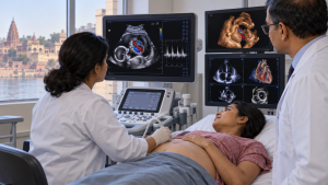
Banaras Hindu University’s Department of Cardiology at the Institute of Medical Sciences inaugurated a fetal echocardiography machine and an advanced cardiac imaging facility in Varanasi on August 11, 2026.
New echocardiography facilities
The fetal echocardiography system is intended to assess the developing heart and help detect congenital heart abnormalities during pregnancy. The advanced laboratory includes a 4D echocardiography system and offers 3D transesophageal and transthoracic echocardiography, strain imaging, stress echocardiography and contrast echocardiography.
Important Facts for Exams
- Doppler assessment: Doppler imaging can show blood flow through the fetal heart, great vessels and umbilical vessels.
- Transoesophageal imaging: Transoesophageal echocardiography places its probe in the oesophagus behind the heart, often producing higher-resolution images than transthoracic imaging.
- Moving 3D images: Four-dimensional echocardiography is three-dimensional imaging with movement in the picture.
Category: Uttar Pradesh
Originally written on
August 11, 2026
and last modified on
November 21, 2026.
| Related | |
|---|---|