
First 3D anatomic heart model printed using multiple imaging techniques
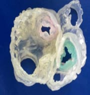
Team of heart experts including an Indian-origin surgeon Dr. Joseph Vettukattil for first time have printed three dimensional (3D) anatomic heart model using multiple imaging techniques.
This hybrid 3D model of heart was successfully printed using integration of 3D transesophageal echocardiography (3DTEE) and computed tomography (CT).
The team had used specialised software to register images in order to selectively integrate datasets and produce an accurate anatomic model of the heart.
This study is considered as a huge leap for individualised medicine in the medical field of cardiology and congenital heart disease. This technological breakthrough will help to promote better diagnostic capability and improve interventional and surgical planning and also provide alternative to surgery in cases of Heart diseases.
This technology also opens the new way in medical technology for producing anatomical parts by using the combination with a third tool of magnetic resonance imaging (MRI).
Dr. Joseph Vettukattil is known internationally for his work and research in the field of three and four-dimensional echocardiography.
Related Posts
Month: Current Affairs - June, 2015
6 Comments
Leave a Reply
You must be logged in to post a comment.
sarthak
June 30, 2015 at 9:41 pmgood
sarthak
June 30, 2015 at 9:41 pmgood
santhiya
July 27, 2015 at 9:07 amgood
santhiya
July 27, 2015 at 9:07 amgood
Ramadevi
April 28, 2016 at 3:23 pmthanks for the information
Ramadevi
April 28, 2016 at 3:23 pmthanks for the information